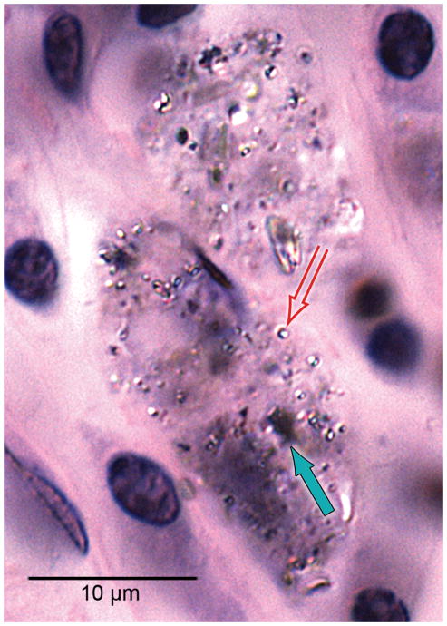
Fig. 4.
Macrophages containing both black particulate material (solid arrow) and birefringent material (hollow arrow) consistent with phagocytized platinum and silicone respectively were commonly found in the fibrous tissue sheath surrounding the cochlear implant electrode, here in case #21 (H&E stain).

