Fig. 6.


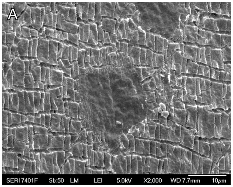
Fig. 6A. Scanning electron microscopy (SEM) of case #27, an individual who had a cochlear implant (Advanced Bionics High Res Helix) implanted two years previously in the right ear. Micro-fragmentation of the silicone carrier was seen.
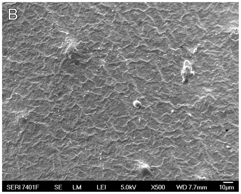
Fig. 6B. SEM of a previously unimplanted control (Advanced Bionics High Res 90 Helix cochlear implant). The silastic carrier did not demonstrate the micro-fragmentation seen in Figure 6A.
