Fig. 7.


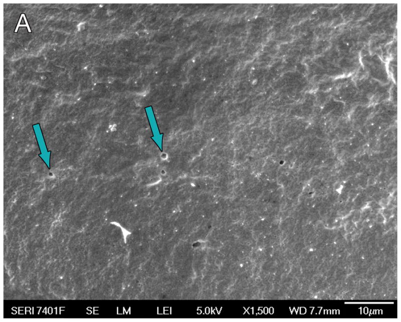
Fig. 7A. Scanning electron microscopy (SEM) of the silastic carrier implanted in case #19 (Nucleus modiolar-hugging cochlear implant) implanted three years prior to death, demonstrated small circular defects (arrows) consistent with loss of material from the silicone carrier.
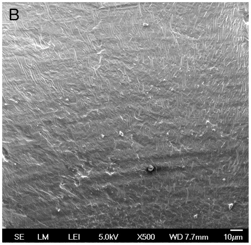
Fig. 7B. Control Nucleus 24 electrode array not previously implanted. The silicone carrier showed no circular defects, as were shown in Figure 7A.
