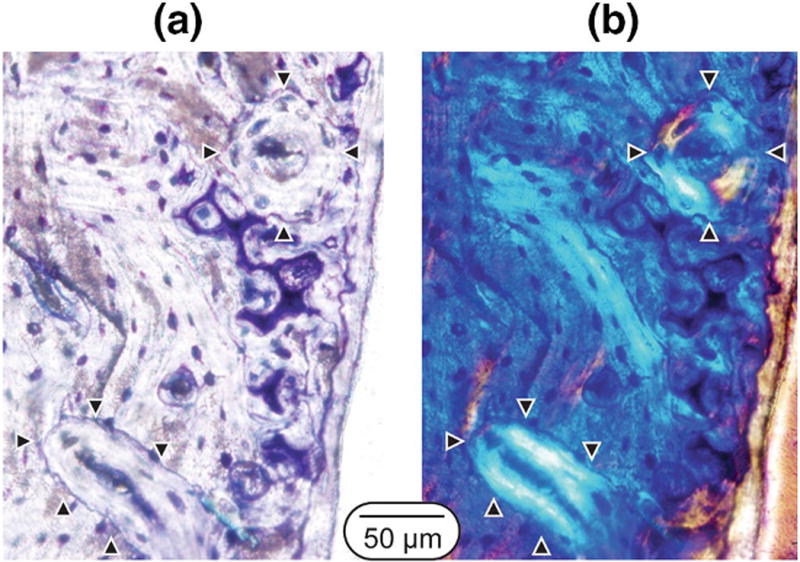
Figure 4.

Example of secondary osteons in a lactation/low Ca rat at the end of the recovery phase. The images are from transverse sections taken at the distal femoral site. (a) Toluidine blue stained image showing cement lines and remnant calcified cartilage within the cortex and (b) matched polarized light images of concentric lamellae. Arrowheads point to secondary osteons within the field of view.
