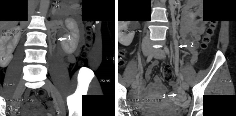
Figure 7.
Computer tomography (CT) scans obtained in patient K.
Notes: The tortuous left accessory ovarian vein (left) and the trunk of the left ovarian vein. The dilated grapelike plexuses and parametrial veins are visualized. 1= additional left gonadal vein; 2= left gonadal vein; 3= the dilated grapelike plexuses and parametrial veins.

