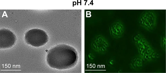
Figure 3.

Characterization of bright-field and fluorescent images at pH 7.4, respectively. Bright-field (A) and fluorescence (B) images of exenatide–PEG-b-(PELG50-g-PLL3) and exenatide–FITC-PEG-b-(PELG50-g-PLL3).
Abbreviations: SEM, scanning electron microscopy; PEG-b-(PELG50-g-PLL3), poly(ethylene glycol)-b-brush poly(l-lysine) polymer; FITC-PEG-b-(PELG-g-PLL), FITC-poly(ethylene glycol)-block-brush poly(l-lysine) polymers.
