
Figure 3.
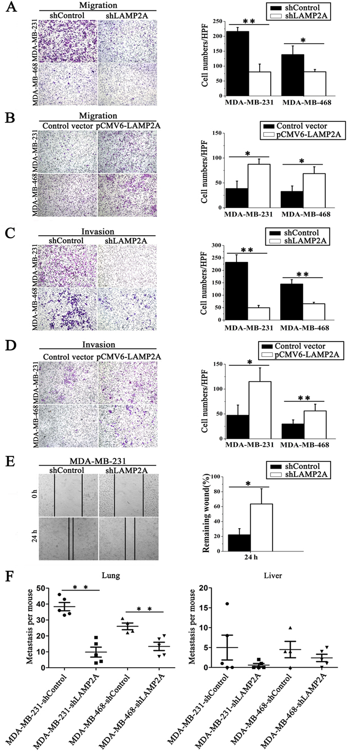
CMA promotes breast cancer cell migration and invasion in vitro and in vivo. (A) and (B) The migration of breast cancer cells was detected using a Transwell migration assay. shLAMP2A or shControl MDA-MB-231 and MDA-MB-468 cells (A) or transient LAMP2A-overexpressing or control MDA-MB-231 and MDA-MB-468 cells (B) were cultured for 6 h, and the cells that migrated to the lower chamber were observed by a microscope after crystal violet staining (left) and counted (right). (C) and (D) The invasion ability of the two breast cancer cell lines was detected using a Transwell Matrigel invasion assay. shLAMP2A or shControl cells (C) and LAMP2A-overexpressing or control cells (D) were cultured for 48 h, and cells that invaded into the lower chamber were observed by a microscope after crystal violet staining (left) and counted (right). (E) The migration of breast cancer cells was measured by a wound-healing assay. The cells were cultured for 24 h after the wound scratch, and the size of the wound at different times was observed by a microscope (left). The size of the wound was quantified (right). All values are the means ± SD of three different experiments; *P < 0.05, **P < 0.01. (F) Nude mice were injected with either shLAMP2A or shControl MDA-MB-231 cells or with shLAMP2A or shControl MDA-MB-468 breast cancer cells via the tail vein. At 60 days after the tail vein injection, the numbers of tumor nodes in the lungs (left) and livers (right) were counted (n = 4 to 5, **P < 0.01, shLAMP2A or shControl).