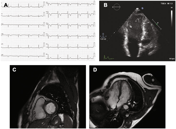
Figure 1.

Non-invasive diagnostic tests in an Arrhythmogenic CardioMyopathy (ACM) patient. (A) Baseline 12-lead ECG shows regular sinus rhythm with inverted T waves in the precordial leads from V1 to V4. (B) Four-chamber echocardiographic imaging shows enlarged right ventricular diastolic volumes. Cardiac magnetic resonance: four-chamber short-axis (C) and long axis (D) images reveal dilated right ventricle with segmental kinesis abnormalities, mainly involving the inferolateral wall.
