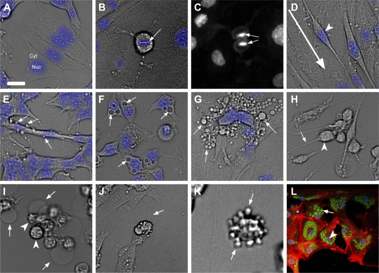
Figure 4.
Cell-based phenotypic screening endpoints for phenotype-based drug discovery.
Notes: (A) Cell morphology endpoint assay setup showing a brightfield image of cells with nuclei stained in blue. (B–C) Cell proliferation endpoint assay showing brightfield images of metaphase cells with metaphase plate (arrow) (B) and nuclei staining of cells in anaphase (arrow) (C). (D) Migrating cells in an adherent fibroblast culture with arrowhead pointing at the nucleus and the direction of migration shown by an arrow. (E) Cell ruffling phenotype showing ruffles on cells (arrows). (F) Phenotype of cells with accumulation of vacuoles (arrows). (G) Morphology screening assay endpoint showing extracellular vesicles (arrows). (H) Cell retraction assay showing rounding cells (arrowhead) and thinning cellular processes (arrow). (I) Necrotic cell assay endpoint showing bulged and broken cytoplasm (arrow) and rounded nucleus (arrowhead). (J) Extracellular projection of cells (arrow) in a morphology-based assay. (K) Cell blebbing (arrow) on rounded up cells. (L) Staining of lipid droplets inside a cell with Bodipy (green), nuclei (arrow head; blue) and tubulin (red). Scale bar: 25 µm; brightfield images of cells and Hoechst-stained nucleus in blue or white in panel (C) are shown.
Abbreviations: Cyt, cytoplasm; Nuc, nucleus.

