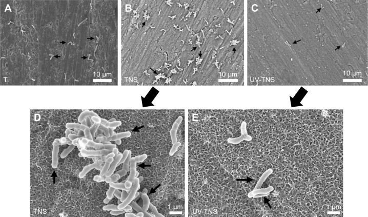
Figure 4.
SEM analysis of the morphology of bacteria attached to sample discs.
Notes: (A) Ti, (B and D) TNS, and (C and E) UV-TNS discs were incubated with A. oris strain MG-1 for 1 hour and then evaluated by SEM. Magnification: 2,000× for (A–C) (scale bar =10 μm) and 10,000× for (D and E) (scale bar =1 μm). A. oris MG-1 displayed a rod-like shape with a complete cell wall on both TNS and UV-TNS; lyzed or irregularly shaped bacteria were not observed.
Abbreviations: A. oris, Actinomyces oris; TNS, titanium with nanonetwork structures; UV-TNS, ultraviolet-treated titanium with nanonetwork structures; SEM, scanning electron microscopy.

