
Figure 2.
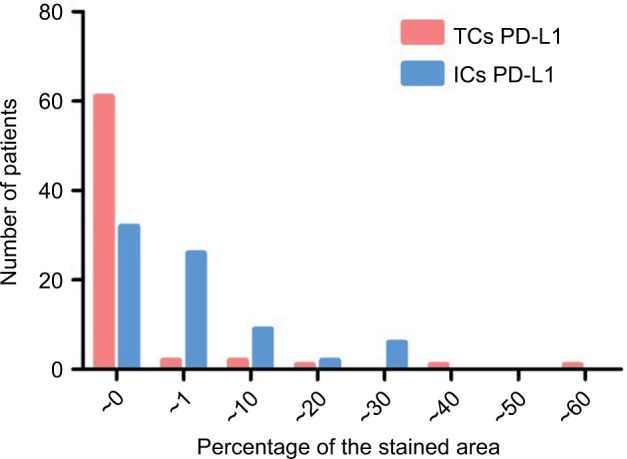
Distribution pattern of levels of PD-L1 expression in TCs and ICs.
Notes: The level of PD-L1 expression was quantified on the basis of percentage of the stained area. PD-L1 expression in TCs was low for most patients, while that in ICs was relatively high.
Abbreviations: PD-L1, programmed death-ligand 1; TC, tumor cell; IC, interstitial cell.