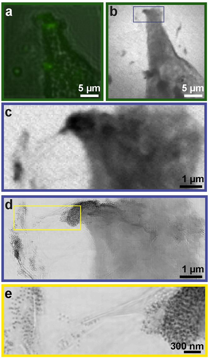
Figure 2.

Localization of functionalized nanoparticles in a cellular context with correlative microscopy. (a) Part of a HeLa cell containing functionalized nanoparticles was first identified using fluorescent microscopy. (b) The same region was imaged using a coarse STXM scan. (c) A fine STXM scan was then performed on a smaller region of interest and a tomographic tilt series was acquired from this region. (d) Ptychographic imaging was performed on the same region as (c) to obtain higher resolution information. (e) Individual nanoparticles within and around the leading edge of the cell identified from the ptychographic reconstruction.
