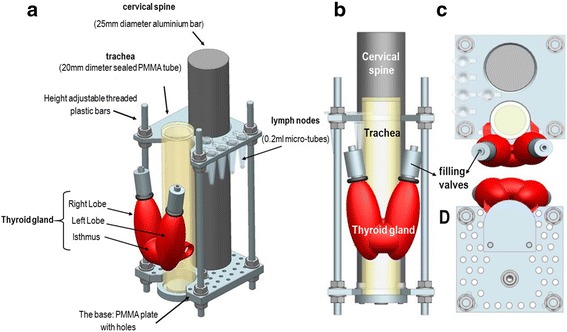
Fig. 2.

Schematics of the internal jig showing the mounted simulated thyroid gland (red) and simulated sentinel lymph nodes (SLNs). The shape and position of the thyroid phantom relative to the simulated trachea: anterolateral (a), anterior (b), superior (c), and inferior (d) views
