Fig. 3.

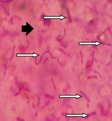
Vertical cross-section and internal appearance of bead at ×100 magnification after gram staining [see the positive gram bacteria (rightwards black arrow) are distributed randomly in the alginate matrix (rightwards white arrow)]

Vertical cross-section and internal appearance of bead at ×100 magnification after gram staining [see the positive gram bacteria (rightwards black arrow) are distributed randomly in the alginate matrix (rightwards white arrow)]