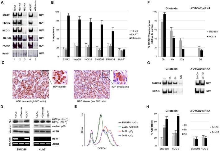
FIGURE 1.
Gliotoxin selectively induces apoptosis in nuclear NOTCH2/CSL active cell lines of solid tumor origin. (A) Electrophoretic mobility shift assays (EMSA) and supershift/interference assays conducted with antibodies specific for N1IC (bTAN 20, N1-Ab) and N2IC (C651.6DbHN, N2-Ab) revealed that NOTCH2 is the dominant active NOTCH family member bound on CSL sites in the indicated cell lines (left panel). Cells were incubated with 5 μM DAPT or 0.2 μM gliotoxin for 1 day and the sensitivity of DNA-bound N2IC complexes to the compounds was determined by EMSA (right panel). (B) Corresponding FACS analysis showing the effect of 5 μM DAPT and 0.2 μM gliotoxin on apoptosis. (C) Immunohistochemistry, showing the nuclear/cytoplasmic localisation of NOTCH2 in HCC tissues in relation to the N/C ratio. (D) Western blotting and RT-PCR showing the effect of gliotoxin (0.2 μM) and DAPT (5 μM) on the expression of NOTCH2 and its target gene HEY1 in nuclear N2IC positive SNU398 and nuclear N2IC negative Huh7 HCC cells after 1 day of incubation. Nuclear NFκB p65, a redox sensitive transcription factor, was not affected by gliotoxin and DAPT. (E) 0.2 μM gliotoxin did not induce oxidative stress in SNU398 cells. Cells were treated with 0.2 μM gliotoxin and with two different concentrations of H2O2 and the ROS concentration was determined by a DCFDA assay via flow cytometry. (F–H) Quantitative RT-PCR (qPCR), EMSA, and FACS comparing the time dependent effect of NOTCH2 inhibition by gliotoxin and siRNA on NOTCH2 mRNA expression, NOTCH2/CSL complexes, and on apoptosis in SNU398 and HCC-3 cells. Data is given as mean from three independent experiments ± standard deviation. ∗The nuclear NOTCH2 negative HCC cell line Huh7 cells served as negative control.

