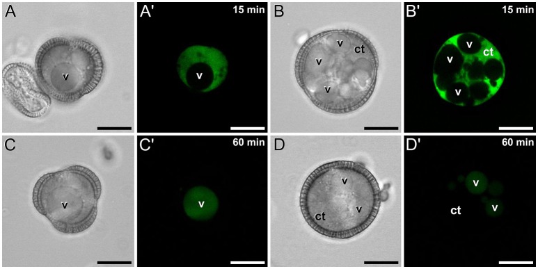
Figure 2.
FF staining of rapeseed microspores and pollen after 15 and 60 min of observation. Phase contrast (A–D) and fluorescence (A′–D′) pairs of pictures are shown. Vacuolate microspore (A,A′) and early binucleated pollen (B,B′) after 15 min of observation. Vacuolate microspore (C,C′) and early binucleated pollen (D,D′) after 60 min of observation. Note the change of FF staining localization from nuclear-cytosolic to vacuolar in both cell types. Ct, cytosol; v, vacuole. Bars: 10 μm.

