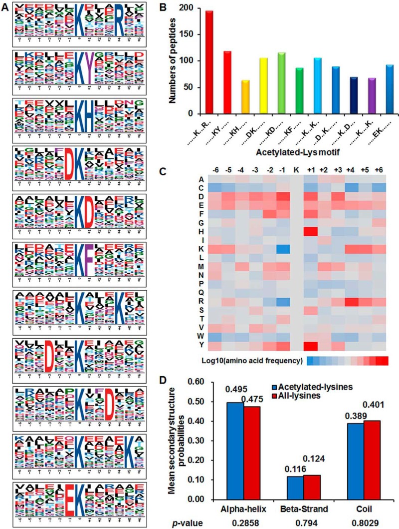
Fig. 2.
Analysis of lysine acetylation sites. A, Probability logos of significantly enriched acetylation site motifs after aligning peptide sequences, including 12 residues surrounding the acetylated lysine residue, using motif-x. B, The number of identified peptides containing acetylated lysine in each motif. C, The relative abundance of amino acid residues flanking the acetylation sites represented by an intensity map. The intensity map shows the relative abundance for ±6 amino acids from the Synechococcus lysine-acetylated site. The colors in the intensity map represent the log10 of the ratio of frequencies within acetyl-13-mers versus nonacetyl-13-mers (red shows enrichment, blue shows depletion). D, Distribution of secondary structures containing lysine acetylation sites. The different secondary structures (α-helix, β-strand and -coil) of acetylated lysine identified in this study were compared with the secondary structures of all lysine identified through proteomics.

