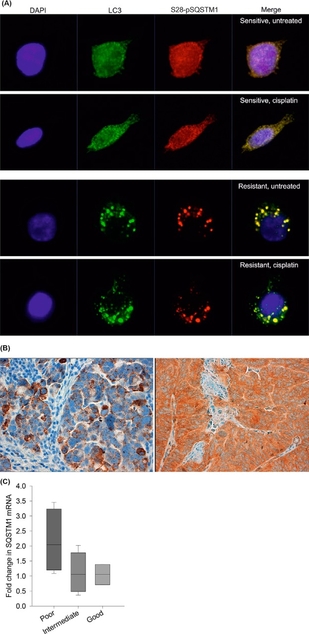
Fig. 9.
Expression and localization of SQSTM1 in cell lines and human tumor specimens. A, Subcellular localization of S28-pSQSTM1 in platinum sensitive M019i and resistant M019iCis cells using immunocytochemical staining. DAPI is represented with blue stain. S28-pSQSTM1 (red) is localized in cytoplasm of the sensitive cell line while it partially localizes into autophagosomes when treated with cisplatin. In resistant cells S28-pSQSTM1 is observed mainly in autophagosomes marked with LC3 staining (green) even without cisplatin treatment. B, Representative SQSTM1 RNA in situ hybridization (left) and immunohistochemical staining (right) shows SQSTM1 expression in the tumor cells but not in surrounding stroma. C, The fold difference in SQSTM1 mRNA expression in eleven HGSOC tumor pairs before and after neoadjuvant chemotherapy classified by histological response to chemotherapy. SQSTM1 mRNA levels (mean, S.E., n = 3) were increased during chemotherapy as presented with the fold difference over 1.0 especially in tumors in which the chemotherapy response was poor. The change was not statistically significant, possibly due to a small number of sample pairs.

