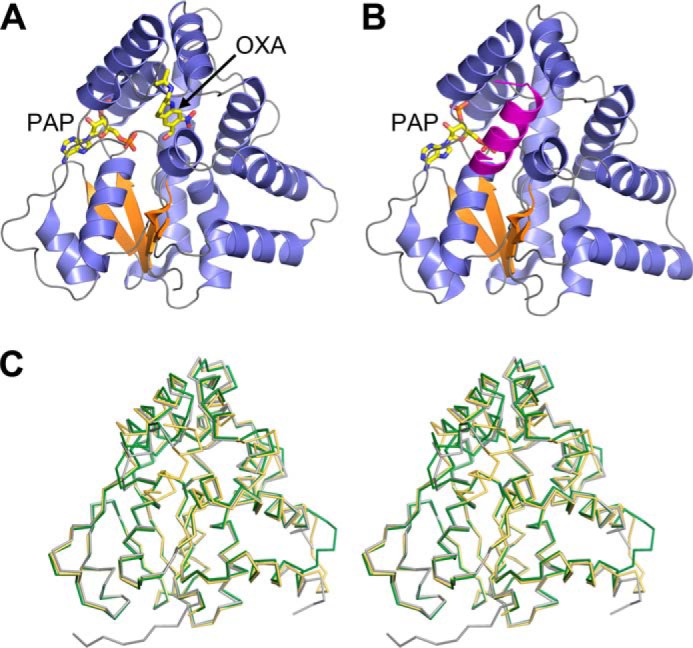
Figure 1.

A, schematic representation of the ShSULT–PAP–oxamniquine (OXA) complex crystal structure. β strands are colored orange, and α helices are colored slate blue. B, schematic representation of the SjSULT–PAP complex crystal structure. Note that the predicted biological assembly is shown with a chain-swapped C-terminal α helix from the crystal symmetry-related molecule colored magenta. For clarity, the C-terminal Gly-Ser His8 affinity tag is not shown. C, walleyed stereoview of the superposition of the Cα traces of ShSULT (dark green) and SjSULT (light orange) onto SmSULT (gray).
