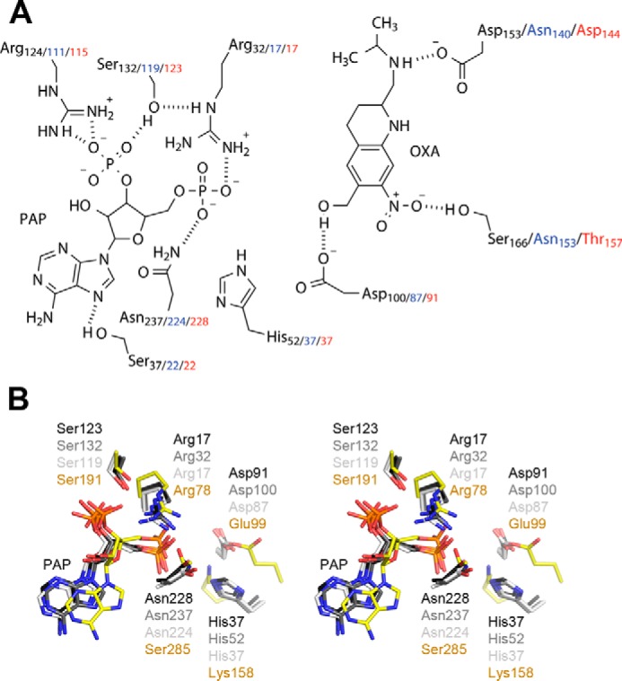
Figure 3.

A, schematic of the ShSULT active site (SjSULT residues in blue and SmSULT residues in red) showing oxamniquine (OXA) in position for sulfate transfer from PAPS (PAP shown). Hydrogen bonds are indicated as dotted lines. B, walleyed stereoview of the superposition of TPST2 and schistosome SULT active-site residues. SmSULT, dark gray; ShSULT, medium gray; SjSULT, light gray; TPST2, yellow.
