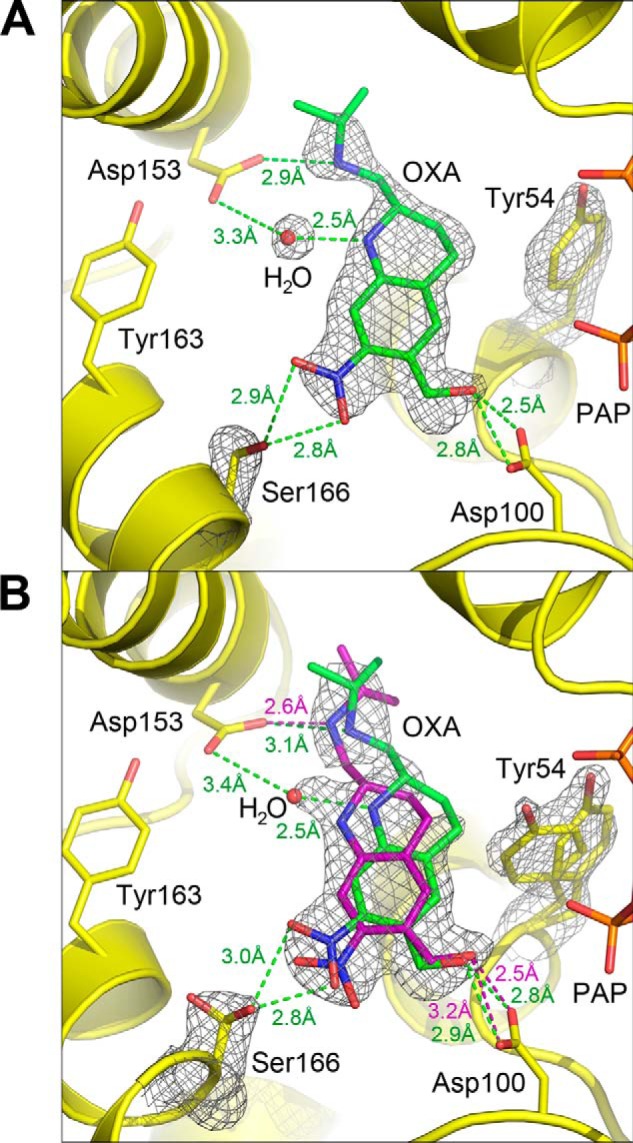
Figure 5.

Crystal structure of ShSULT soaked with racemic oxamniquine. A, chain B shows a single conformation for bound (S)-oxamniquine. A composite omit map (2mFo − DFc) contoured at 1.0 σ is superimposed on the atoms shown. B, oxamniquine (OXA) binds in the ShSULT active site in two positions in chain A determined by the alternate conformations of Tyr-54 and Ser-166. A composite omit map (2mFo − DFc) contoured at 1.0 σ is superimposed on the atoms shown. Oxamniquine alternate confirmations and corresponding hydrogen bonding interactions are indicated in green and purple. A solvent atom is positioned between Asp-153 and oxamniquine when the alternate conformation shown in green is present (compare with A).
