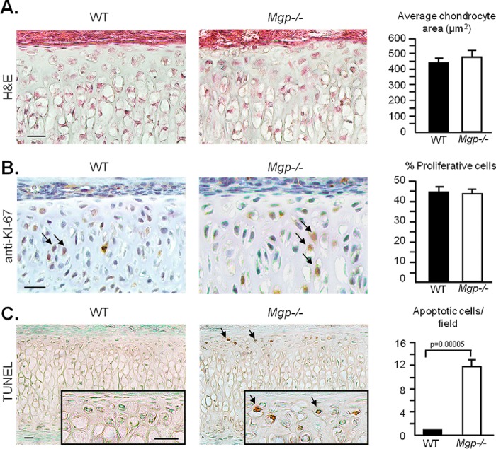
Figure 7.
MGP-deficient septal chondrocytes undergo apoptosis. A, quantification of cell area on histological sections of WT and Mgp−/− nasal septa stained with H&E shows no difference in chondrocyte size between the genotypes. Three fields were quantified per sample (n = 3 in both groups). Scale bar = 20 μm. B, anti-Ki67 antibody and hematoxylin staining of septal sections showing the proliferating chondrocytes (arrows). Quantification of proliferative cells/total cell count shows no difference between WT and Mgp−/− mice (n = 6 in both groups). Scale bar = 20 μm. C, colorimetric apoptosis detection assay (TUNEL) performed on WT and Mgp−/− septal sections shows the presence of apoptotic cells (arrows) in the MGP-deficient nasal septum but not in the WT nasal septum sections. The sections were counterstained with methyl green (n = 3 in both groups). Scale bars = 20 μm. All analyses were performed on 2-week-old mice. Statistical analysis: Student's t test in all cases.

