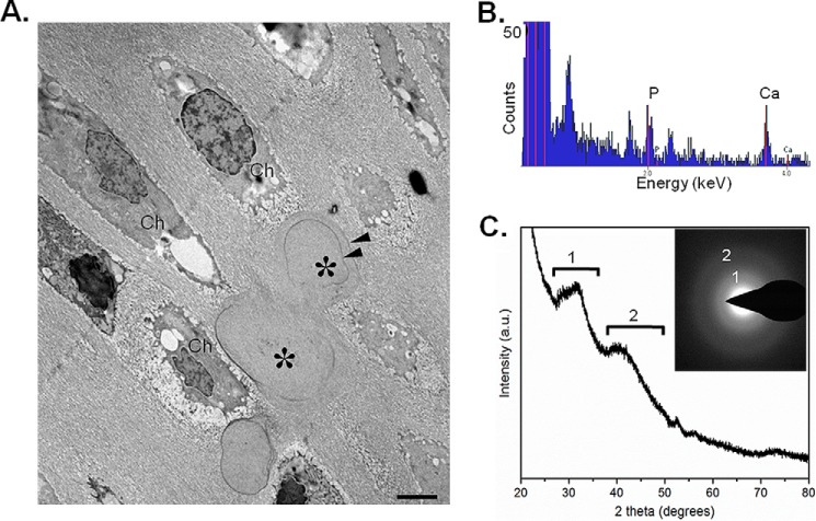
Figure 9.
Amorphous calcium phosphate as the main mineral species in MGP-deficient nasal septum. A, transmission electron microscopy of 1-week-old Mgp−/− nasal septum showing the presence of chondrocytes (Ch) and mineral deposits (asterisks) having an unusual globular shape. Note the incremental lines (suggesting periodic mineral deposition) within the mineral deposits (arrowheads). B, energy-dispersive X-ray spectroscopy showing the presence of phosphorus and calcium within the mineral deposits. C, X-ray diffraction showing that the mineral phase is largely amorphous calcium phosphate. Minor spectral peaks labeled 1 and 2 indicate tendencies toward crystallization (initiation) of an apatitic phase. a.u., arbitrary units. Inset, electron diffraction pattern confirming the presence of mostly amorphous mineral showing only diffuse (rather than sharp) electron diffraction rings labeled 1 and 2 (n = 3 mice for each group and each experiment).

