Figure 2.
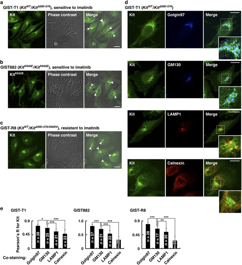
In GIST cell lines, Kit(mut) localizes preferentially on the Golgi apparatus. (a–c) GIST-T1 (a), GIST882 (b) and GIST-R8 (c) were immuno-stained with anti-Kit. Phase contrast images are shown. Arrowheads indicate the perinuclear region. Bars, 20 μm. (d) GIST-T1 cells were double-stained with anti-Kit plus the indicated antibody. Insets show magnified images of the boxed areas. Bars, 20 μm. Golgin97 (trans-Golgi marker), blue; GM130, (cis-Golgi marker), blue; calnexin (ER marker), red; LAMP1 (lysosome-associated membrane protein 1, endo/lysosome marker), red. (e) Pearson’s R correlation coefficients were calculated by intensity analysis of Kit vs organelle markers. Results are means±s.d. (n=17–31). *P<0.05, **P<0.01, ***P<0.001.

