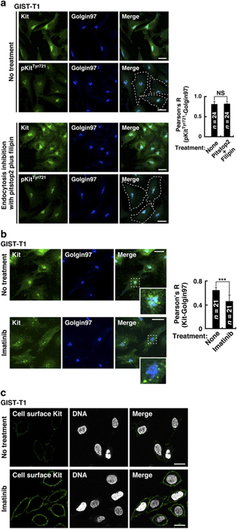
Figure 4.
Kit(mut) accumulates on the Golgi during the early secretory pathway but not after endocytosis in a manner that depends on its kinase activity. (a) GIST-T1 cells were treated with 50 μM pitstop2 plus 1 μg/ml filipin for 24 h to block endocytosis, then immuno-stained with anti-Kit (green), anti-pKitTyr721 (green) and anti-golgin97 (Golgi marker, green). Dashed lines indicate cell borders. Bars, 20 μm. The graph shows Pearson’s R between pKitTyr721 and golgin97. Results are means±s.d. (n=24). NS, not significant. P=0.68. (b, c) GIST-T1 cells were treated with 200 nM imatinib (Kit inhibitor) for 4 h. (b) Cells were stained for Kit (green) and golgin97 (blue). Insets show magnified images of the boxed areas. Bars, 20 μm. The graph shows Pearson’s R between Kit and golgin97. Results are means±s.d. (n=21). ***P<0.001. (c) Cells stained for the Kit extracellular domain (cell surface Kit, green) and DNA (white) without permeabilization. Bars, 20 μm.

