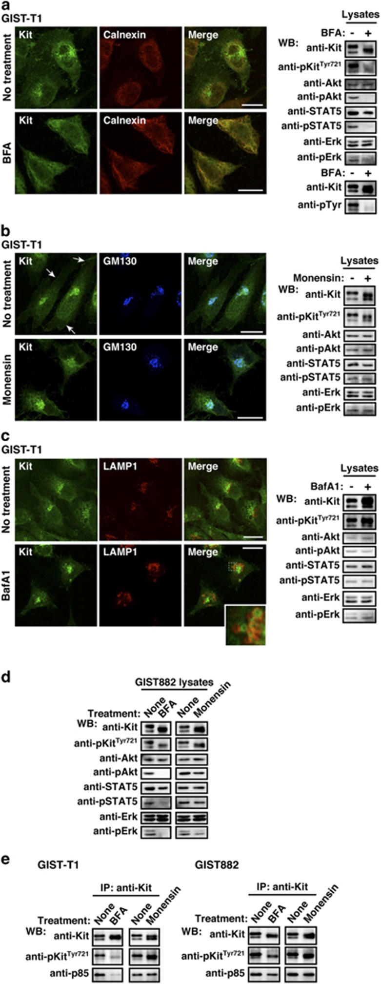
Figure 5.
Kit(mut) on the Golgi signals and activates the PI3K-Akt pathway, STAT5, and Erk. (a–c) GIST-T1 cells were treated with (a) 1 μM BFA (brefeldin A; blocks ER export to the Golgi) for 16 h, (b) 250 nM monensin (blocks Golgi export) for 24 h, or (c) 100 nM bafilomycin A1 (BafA1; blocks endo/lysosomal trafficking) for 24 h. Cells were stained with anti-Kit (green) in conjunction with anti-calnexin (ER marker, red), anti-GM130 (Golgi marker, blue), or anti-LAMP1 (endo/lysosome marker, red). Arrows indicate the PM region. An inset shows a magnified image of the boxed area. Bars, 20 μm. Immunoblots are shown. Phosphorylated proteins are presented as pKit, pAkt, pSTAT5, and pErk. (d) GIST882 cells were treated with 1 μM BFA for 16 h or 250 nM monensin for 24 h. Lysates were immunoblotted. (e) GIST-T1 and GIST882 were treated with 1 μM BFA for 16 h or 250 nM monensin for 24 h. Anti-Kit immunoprecipitates were immunoblotted.

