. 2015 May 19;6(8):4636–4642. doi: 10.1039/c5sc00952a

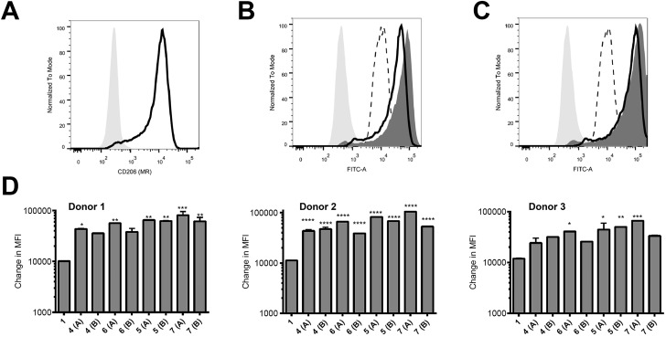
Fig. 2. Increased targeting of glycosylated peptides to MR-positive APC. (A) Expression levels of CD206 (MR) on the APC were determined using an anti-CD206 antibody. (B and C) APC were treated with 10 μM of peptides 1 and 4–7 at 37 °C for 60 minutes and analysed by flow cytometry. Results in (B) show the binding for a representative donor of 1 (dotted line) compared to 4 (black line) and 6 (dark grey histogram) relative to the unstained sample (light grey shaded histogram). Results in (C) show the binding of 1 (dotted line) compared to 5 (black line) and 7 (dark grey histogram) relative to the unstained sample (light grey shaded histogram). In (D), results are given as the change in measured fluorescence intensity (MFI, calculated as the MFI of sample minus the MFI of the untreated control); each sample was tested in duplicate. Monocytes cultured from three individual donors are shown. Statistical significance for each sample (compared to 1) was calculated using a one way Anova followed by Dunnett's multiple comparison test; *p < 0.05, **p < 0.01, ***p < 0.001.

Fig. 2