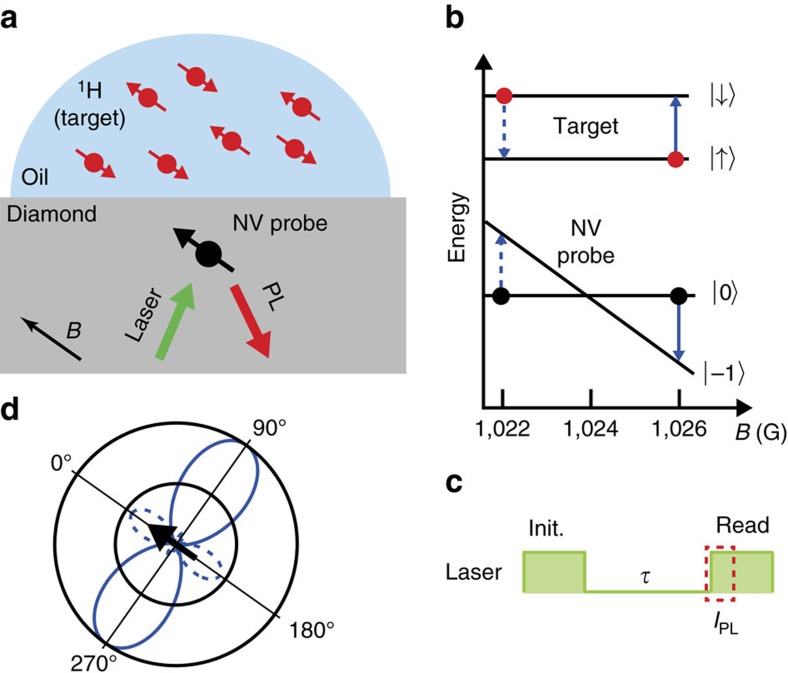
Figure 1. Principle of microwave-free nano-nuclear magnetic resonance.
(a) Schematic of the experimental setup. The protons within an organic sample external to the diamond are the targets probed by a shallow nitrogen-vacancy (NV) centre. A 532 nm green laser is used to initialize the NV spin while the red photoluminescence (PL) is measured via a single-photon detector. The background magnetic field is aligned with the NV quantization axis with a variable strength, B. (b) Schematic of the energy levels of the NV (spin states |0〉 and |−1〉, neglecting the hyperfine structure for clarity) and of a target nuclear spin such as 1H (states |↑〉 and |↓〉). As B is swept across the ground state level anti-crossing (GSLAC), two resonances can in principle occur where the NV and target spins can exchange energy via their mutual magnetic dipole–dipole interaction, causing an increase in their respective longitudinal relaxation rate. (c) The measurement sequence consists of laser pulses to initialize and subsequently read out the NV spin state, separated by a wait time τ. (d) Polar plot of the relaxation rate induced by a single spin, Γ1,ext, as a function of the angle θ between the quantization axis and the NV-target separation. The solid (dashed) line corresponds to the after-GSLAC (before-GSLAC) resonance. The values are normalized by the global maximum, so that the outer circle corresponds to maximum strength. The polar plot is rotated by θ=54.7°, which is the angle between an NV below a (100) diamond surface, and the closest target spin on the surface.

