
Figure 4.
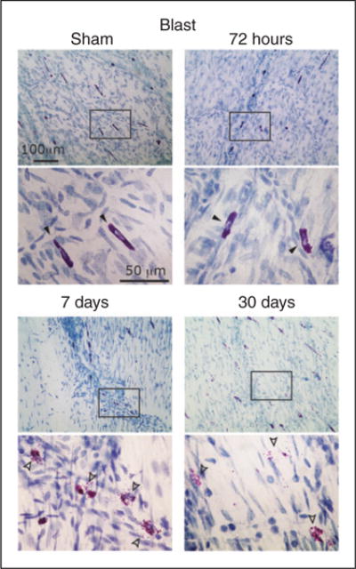
Degranulation of dural MCs following a blast-evoked closed head trauma. Images are taken from samples obtained from the dura outlining the right cortical hemisphere. Note the delayed degranulating effect, first seen at seven days. Also note the robust degranulating effect at 30 days, evidenced by cells with reduced TB staining. Closed arrowheads (non-degranulated MCs) and open arrowheads (degranulated MCs) in the lower magnification point mark the cells shown in the higher magnification images below.