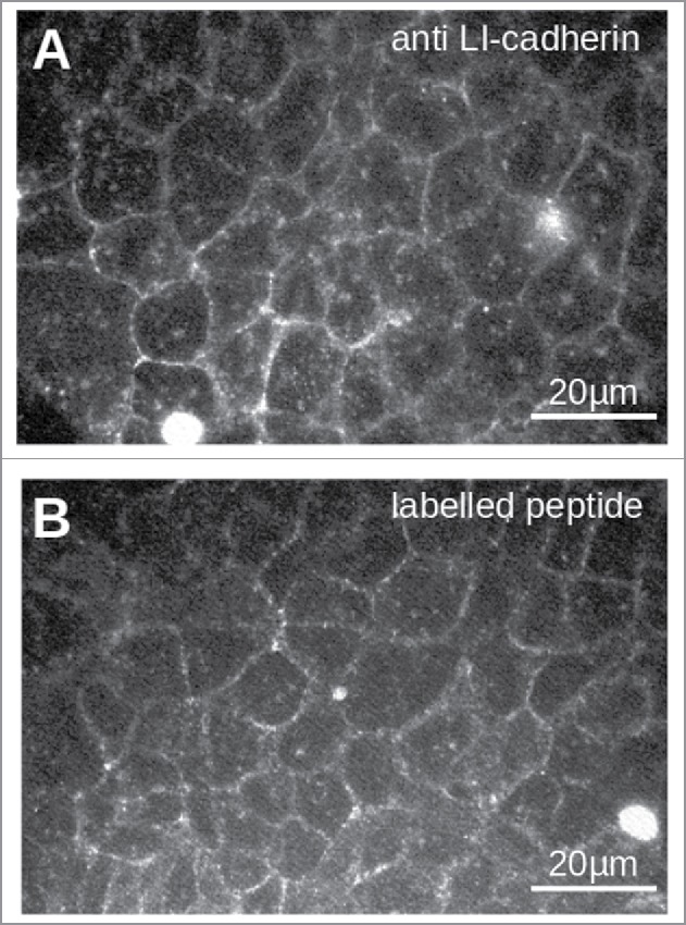
Figure 3.

Staining of LI-cadherin in CACO2-cells with LI-cadherin specific antibodies (A) or with the fluorescently labeled peptide VAALD (B). Clearly the LI-cadherin is mainly found at cell-cell-boarders. Clearly there is a localization of the peptide and the antibody along the cell boarders. It has to be emphasized that the cells for the peptide staining were not permeabilised to see whether or not the peptide can enter the lateral intercellular cleft for our transport measurements.
