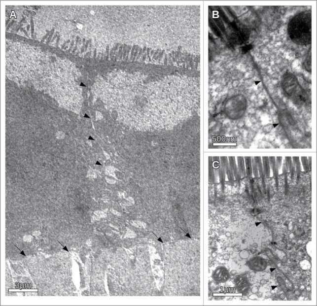
Figure 6.

Transmission electron microscopic images of CACO-cells under different osmotic conditions in the presence and absence of the inhibitory peptide. (A) Overview of the intercellular cleft in the presence of the inhibitory peptide under hypertonic conditions. Apically the microvilli could be seen. The basal membrane on the transwell filter is indicated by arrows. The intercellular cleft (indicated by arrowheads) clearly becomes more and more impaired from apical to basal. Irregular widening of the cleft was observed. (B) Apical section of CACO-cells in the vicinity of the junctional complex under hypertonic conditions. Note no obvious morphological change of the junctional complex. (C) CACO-cells in the absence of the inhibitory peptide under hypertonic conditions. The intercellular cleft (arrow heads) is constantly narrow.
