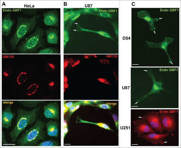
Figure 1.
Dual Golgi and PM localization of GBF1 in GBM cells. HeLa (A) and U87 (B) cells were probed by double label IF with polyclonal rabbit anti-GBF1 and monoclonal mouse anti-GM130 antibodies. GBF1 co-localizes with GM130 to the Golgi, but is also present at tips of protrusions in U87 cells (arrows). (C) D54, U87 and U251 cells were processed for IF with monoclonal mouse anti-GBF1 antibodies. GBF1 localizes to the peri-nuclear Golgi and at tips of protrusions and the leading edge (arrows). Representative images from more than 3 independent experiments. Bar is 10 μm.

