
Figure 1. Whole Body 18F-FDG PET/MR of a patient with Hodgkin Lymphoma.
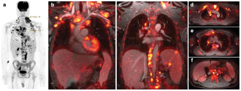
(a) 18F-FDG PET maximum intensity projection shows FDG-avid disease of the neck, chest, axillaries, abdomen and pelvis. (b) Coronal 18F-FDG PET images superimposed on ferumoxytol-enhanced T1-weighted LAVA images clearly show the relation between vessels and multiple hypermetabolic lymph nodes. Three lesions in the spleen are also noted. (c) Axial integrated 18F-FDG PET/LAVA images provide diagnostic information similar to a PET/CT scan, but with improved soft tissue contrast.