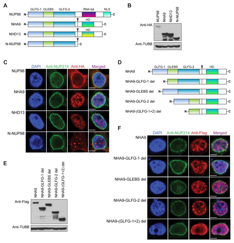
Figure 1. The subcellular localization and expression of NUP98 and NUP98 fusions.
(A) Schematic representation of WT full-length NUP98, NUP98-HOXA9 (NHA9), NUP98-HOXD13 (NHD13) and N-NUP98. Fusion break points are indicated with arrows. (B) Western blot analysis of whole cell lysates from transfected 293T cells detected by anti-HA and anti-beta Tubulin (TUBB) antibodies. One representative experiment of three is shown.
(C) Immunofluorescence staining of 293T cells transfected with the Human influenza hemagglutinin (HA)-tagged full-length NUP98, NHA9, NHD13 and N-NUP98 vectors. Cells were stained with an anti-HA and an anti-NUP214 antibody for double-immunofluorescence analysis by confocal microscopy. HA was labeled with Alexa Fluor 568-conjugated secondary antibody (red), and the NUP214 was labeled with Alexa Fluor 488-conjugated secondary antibody (green). Nuclei were counterstained with DAPI (blue). (D) Schematic of the various NHA9 mutant constructs used in this study. (E) Western blot analysis of whole cell lysates from transfected 293T cells detected by anti-Flag and anti-beta Tubulin (TUBB) antibodies. One representative experiment of three is shown.
(F) Immunofluorescence staining of 293T cells transfected with Flag-Avi-tagged NHA9 and NHA9 mutant vectors. Cells were stained with an anti-Flag and an anti-NUP214 antibody for double-immunofluorescence analysis as described in 1C. Flag was labeled with Alexa Fluor 568-conjugated secondary antibody (red).

