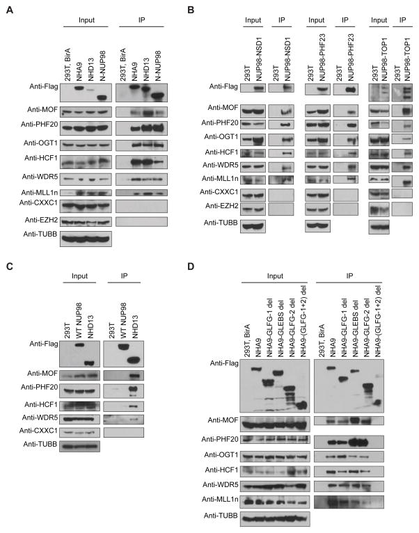
Figure 2. NUP98 fusions interact with MLL1 and the NSL histone-modifying complex.
(A) The Flag-Avi-tagged NHA9, NHD13 and N-NUP98 proteins were stably expressed in 293T cells with co-expression of the BirA biotin ligase. Whole cell lysates were subjected to immunoprecipitation (IP) using streptavidin magnetic beads. Cell lysates (Input) and proteins eluted from the streptavidin beads (IP) were analyzed by Western blotting with anti-Flag, anti-MOF, anti-PHF20, anti-OGT1, anti-HCF1, anti-WDR5, anti-MLL1n, anti-CXXC1, anti-EZH2 and anti-beta Tubulin (TUBB) antibodies. (B) Co-IP was performed on lysates from Flag-tagged NUP98-NSD1 (left), NUP98-PHF23 (middle) and NUP98-TOP1 (right) transfected 293T cells with an anti-Flag antibody, and analyzed by Western blotting. (C) Immunoblots of Co-IP on lysates from Flag-tagged full-length NUP98 or NHD13 transfected 293T cells with an anti-Flag antibody. (D) Co-IP was performed on lysates from Flag-Avi-tagged NHA9 or NHA9-mutant vectors transfected 293T cells with co-expression of the BirA biotin ligase, and analyzed by Western blotting. Data are representative of three individual experiments. See also Figure S1.

