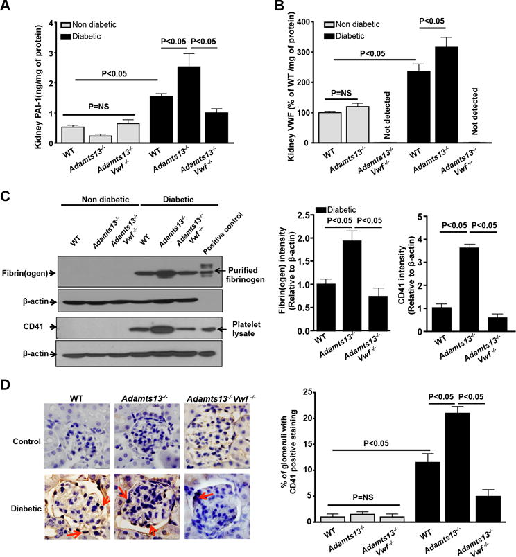
Figure 3. Increased intrarenal thrombosis in Adamts13−/− diabetic mice was VWF-dependent.

Kidney homogenates were prepared after 26 weeks of diabetes induction and as described in methods. A, Kidney PAI-1 levels. B, Kidney VWF levels. C, Left panels show representative images of immunoblots for fibrin(ogen) and CD41 positive platelets accumulation in the kidney homogenates of diabetic mice, but not in non-diabetic mice. Purified fibrin(ogen) and mouse platelet lysate was used as positive control. b-actin was used as loading control. Right panel shows densitometric quantification of the fibrin(ogen) and CD41 immunoblots (C) normalized to corresponding b-actin (loading control). D. Left panel shows representative images of CD41 positive stained microthrombi (red arrows) in the kidney section and right panel shows quantification. Data are presented as mean ± SEM. N=5 mice/group. P<0.05 for diabetic mice when compared to non-diabetic mice in all the groups. Statistical analysis: one-way ANOVA followed by Holm-Sidak multiple comparison test.
