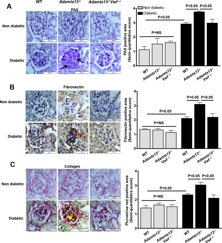
Figure 4. Increased mesangial cell expansion and extracellular matrix deposition in the glomeruli in Adamts13−/− diabetic mice was VWF-dependent.

Left panel shows representative images of A, PAS stained (black arrow) kidney section, B, fibronectin stained (red arrow) kidney section and C, picrosirus red (yellow arrow) stained kidney section. Right panel shows semi-quantitative score for the respective staining. P<0.05 for diabetic mice when compared to non-diabetic mice in all the groups. Data are presented as mean ± SEM. Mean score of 5–20 glomeruli from each mouse were taken from 5–6 mice/group. Statistical analysis: one-way ANOVA followed by Holm-Sidak multiple comparison test.
