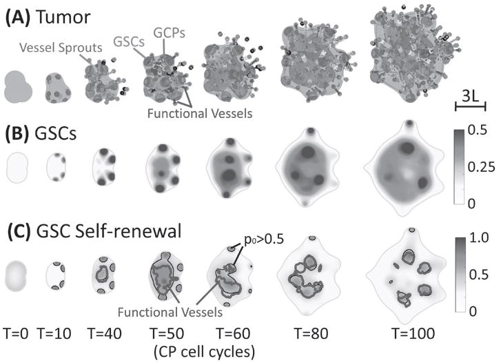
Fig. 1.

VEC-GSC crosstalk promotes GSC proliferation and self-renewal. (A) Time evolution of tumor (φT = 0.5 surface, blue), GSCs (φGSC = 0.3 surface, red), GCPs (φGCP = 0.25 surface, green) and neovasculature (red dots: sprout initiation points; grey: sprouts, blue: functional vessels). At early stages, GSC clusters form near the tumor boundary and fingers develop. Angiogenesis starts around T =39, after which a large GSC cluster forms at the center of tumor due to VEC-GSC crosstalk. (B) 2D slices of GSCs (at z =-1). At T=5, GSC clusters begin to emerge near tumor boundary. (C) 2D slices of the GSC self-renewal probability p (at z=-1) 0 shown together with the p0 = 0.5 (black) and functional vessel density ρFV = 20 (blue) FV contours. In the tumor interior, the functional vessels are colocalized with GSC clusters with p0 > 0.5.
