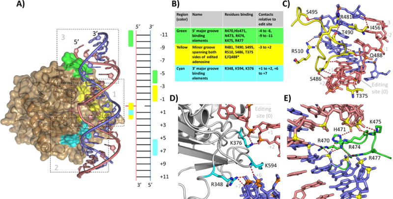
Figure 2.

Interactions of hADAR2d with dsRNA beyond the active site. A: Overview of hADAR2d-RNA structure (5ED1, hADAR2d E488Q with Bdf2 derived 23-mer 8-azanebularane containing RNA) showing three main regions of contact. Edited strand in salmon, complementary stand in blue. Region 1 residues highlighted in yellow, region 2 in cyan, region 3 in green. Ladder diagram shows a secondary structure representation of the protein RNA contact interface (editing site shown in red flipped from the helix) B: Table summarizing the protein residues of each region and the RNA registers bound by each. C: Detail view of region 1. D. Detail view of region 2 (NOTE Fig 2C shows 5ED1 hADAR2d E488Q. E: Detail view of region 3 (NOTE Fig. 2E is from 5ED2, crystal structure of hADAR2D E488Q with hGli1 derived 23mer 8-azanebularne containing RNA. This RNA extends 1 bp farther in the 5′ direction than the Bdf2 derived RNA); (5ED1 and 5ED2 [22]).
