
Figure 2.
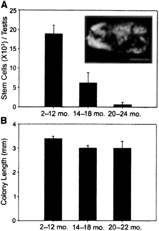
Stem cell activity in mouse testes. (A, inset): Macroscopic appearance of recipient testis transplanted with donor testis cell. Individual blue tubules indicate colonies of spermatogenesis arising from donor spermatogonial stem cells (SSCs). The SSC transplantation assay was used to evaluate stem cell activity in the testes of young (2–12 months), aging (14–18 months), and old (20–24 months) mice. Donor-derived colonies of spermatogenesis are unequivocally identified in recipient seminiferous tubules because they express a reporter gene (i.e., lacZ). Stem cell number (colony number) and kinetics of colony expansion (e.g., colony length) can be evaluated. Bar = 2 mm. (A, graph): Testis stem cell content for young (18.9 ± 2.3 ×10³, n = 12 experiments, 145 recipient testes), aging (6.1 ± 2.6 ×10³, n = 7 experiments, 84 recipient testes), and old (0.7 ± 0.6 7×10³, n = 6 experiments, 68 recipient testes) ROSA26 mice (young vs, aging, p = .002; young vs. old, p = .001; aging vs. old, p = .329). Total stem cell number per donor testis = colonies per 105 cells transplanted × total cells recovered per donor testis 7 × 20 to account for transplantation efficiency [30]. (B): Colony length (mm) from donor testis cells of young (3.4 ± 0.1, n = 24 recipients, 248 colonies), aging (3.0 ± 0.1, n = 12 recipients, 120 colonies), and old (3.0 ± 0.3, n = 10 recipients, 128 colonies) males was determined in recipients by using a dissecting microscope and digital imaging system (young vs. aging, p = .111; young vs. old, p = .089; aging vs. old, p = .977). No stem cell activity was recovered from testes of 24-month-old males. Bars are mean ± SEM