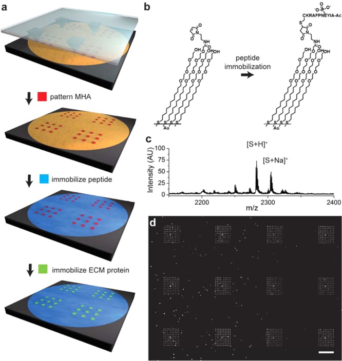
Figure 2.

Nanoarrays were prepared by using PPL to pattern mercaptohexadecanoic acid (MHA) on a gold-coated surface in many 10 × 10 arrays where each spot was 750 nm in diameter and where neighboring spots had a center-to-center spacing of 4.4 μm (a). The remaining areas of gold were then modified with a monolayer presenting maleimide groups against a background of tri(ethylene glycol) groups and used to immobilize a cysteine terminated phosphopeptide (b). The surface was then treated with a solution of fibronectin to allow the adsorption of the extracellular matrix protein to the MHA nanoarray. A SAMDI spectrum of the monolayer confirms immobilization of the peptide (c). The fluorescence micrograph shows fibronectin patterned nanorrays stained with mouse antifibronectin antibody and AlexaFluor568-conjugated goat anti-mouse IgG (d). The scale bar is 40 μm.
