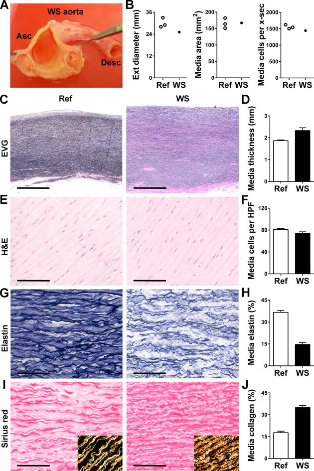
Figure 5. Analysis of Human WS and Referent Ascending Aortas.

(A) Gross examination of the ascending (Asc) aorta from an adult with WS opened to reveal uniformly thickened wall without discrete supravalvular stenosis and rudimentary membranes at the sinotubular junction above the coronary ostia. (B) Measurements of external (ext) diameter, medial area, and total number of medial cells per cross-section (x-sec) in WS specimen and 3 referent (ref) ascending aortas from age- and sex-matched subjects. (C) Representative images of EVG-stained cross-sections (bars: 1 mm). (D) Medial thickness measured at 4 separate points. (E) Representative images of H&E stain (bars: 100 μm). (F) Medial cell density determined in 9 high power fields (HPF). (G) Representative images of elastin stain (bars: 100 μm). (H) Percent of media staining positive for elastin in 9 high power fields. (I) Representative images of sirius red stain (bars: 100 μm) with polarized light images in insets confirming collagen detection. (J) Percent of media staining positive for collagen in 9 high power fields. Data represent mean ± SEM; n = 4–9 replicates from 1 WS specimen vs. 3 referent ascending aortas.
