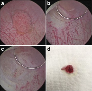
Fig. 1.

a A 1.5-cm-diameter bladder tumor on the right bladder wall. b Macroscopic normal mucosa about 0.5 cm away from the tumor base was margined. Then, the bladder mucosa was subsequently cut in a “flash-firing” fashion. c After the deep muscle layer was reached when normal glistening yellow fat is seen between muscle layers, the loop was moved forward along the muscle layer. d The tumor was resected in one piece
