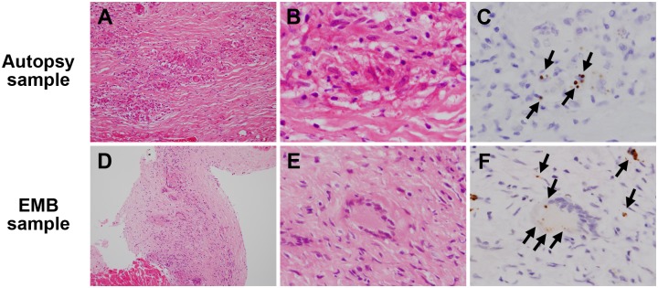
Fig 1. Representative samples with sarcoid granulomas.
An autopsy sample (A–C) and an EMB sample (D–F) from patients with sarcoidosis were stained with haematoxylin and eosin and immunostained with anti-P. acnes antibody. Many sarcoid granulomas were observed at the lower magnification (A, D). Small round bodies indicated by the black arrows (C, F) were found in some of epithelioid cells and multinucleated giant cells of these sarcoid granulomas by immunohistochemistry with anti-P. acnes antibody. Original magnification; ×200 (left), ×1000 (middle and right).

