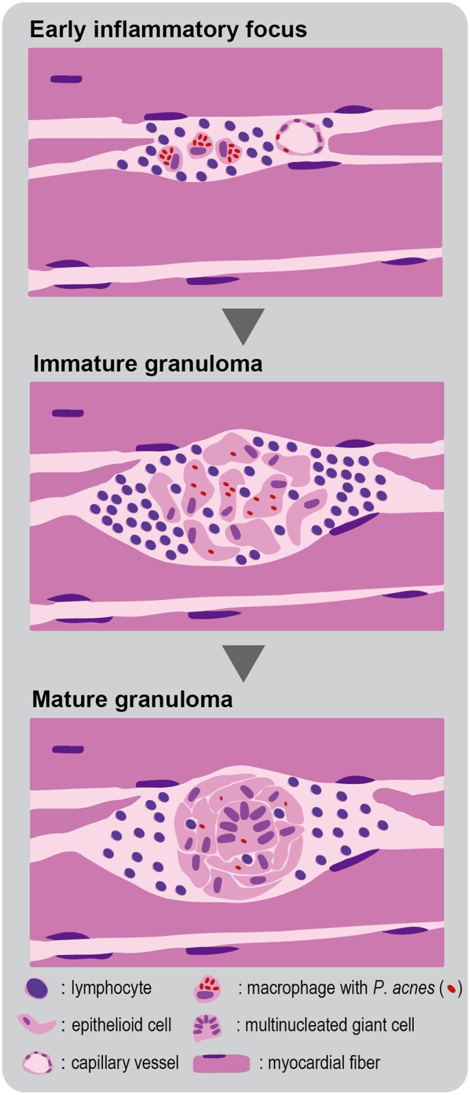
Fig 4. Schematic representation of granulomatous inflammation caused by P. acnes.

Granulomas begin as small collections of lymphocytes and macrophages with intracellular P. acnes (early inflammatory foci) as observed in the minimal or massive inflammatory foci of the CS-group samples. Macrophages change to epithelioid cells and become organized into a cluster of cells (immature granuloma). Further progression results in ball-like clusters of cells and fusion of macrophages into giant cells with or without remaining intracellular P. acnes (mature granuloma).
