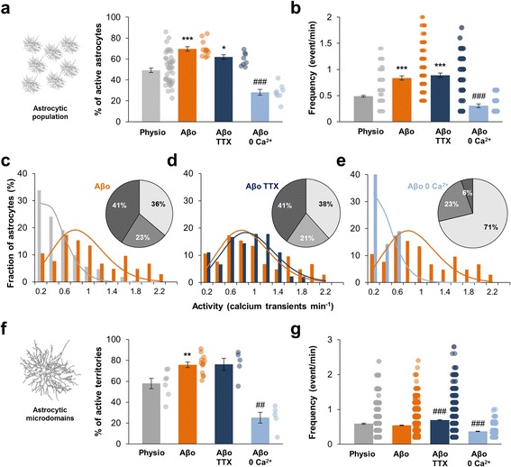
Fig. 3.

Extracellular Aβo application induces astrocytes hyperactivity. a, b. Within the global astrocytic population, proportion of astrocytes displaying calcium activity and frequency of astrocyte calcium activity in physiological condition (grey; n = 43), under 100 nM Aβo application (orange; n = 12), 100 nM Aβo + 500 nM TTX co-application (dark blue; n = 8) and 100 nM Aβo in Ca2+-free medium application (0 Ca2+; light blue; n = 8). c, d, e. Frequency histogram revealing that significantly more astrocytes were hyperactive and less were inactive under 100 nM Aβo application or 100 nM Aβo + 500 nM TTX co-application. Aβo-induced hyperactivity is abolished in Ca2+-free medium. Related theoretical Poisson distributions were shown accordingly. f, g. Within single astrocyte arbor, proportion of subregions displaying calcium activity and frequency of astrocyte calcium activity in physiological condition (grey; n = 7), under 100 nM Aβo application (orange; n = 13), 100 nM Aβo + 500 nM TTX co-application (dark blue; n = 5) and 100 nM Aβo in Ca2+-free medium application (0 Ca2+; light blue; n = 5). Results are compared with the physiological condition with *, p < 0.05; **, p < 0.01 and ***, p < 0.001 or the Aβo condition with #, p < 0.05; ##, p < 0.01 and ###, p < 0.001
