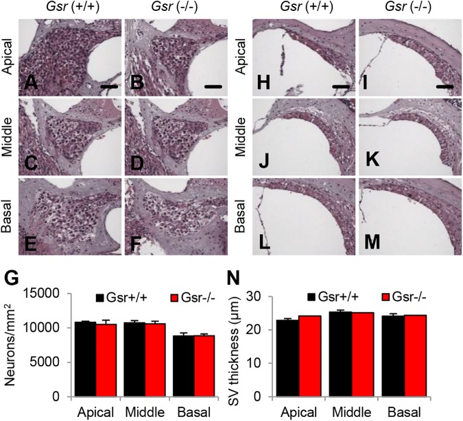
Fig 6. Histological analysis of cochlear spiral ganglion neuron density and stria vascularis thickness in young Gsr+/+ and Gsr-/- mice.
(A-G) The densities of spiral ganglion neurons (SGN) in the apical, middle, and basal regions of cochlear tissues from 3–5 month-old male Gsr+/+ and Gsr-/- mice (N = 4–5) were counted and quantified (G). (H-N) The thickness of stria vascularis (SV) in the apical, middle, and basal region of cochlear tissues from 3–5 month-old Gsr+/+ and Gsr-/- males (N = 4–5) was measured (N). Data are shown as means ±SEM. Scale bar = 25 μm.

