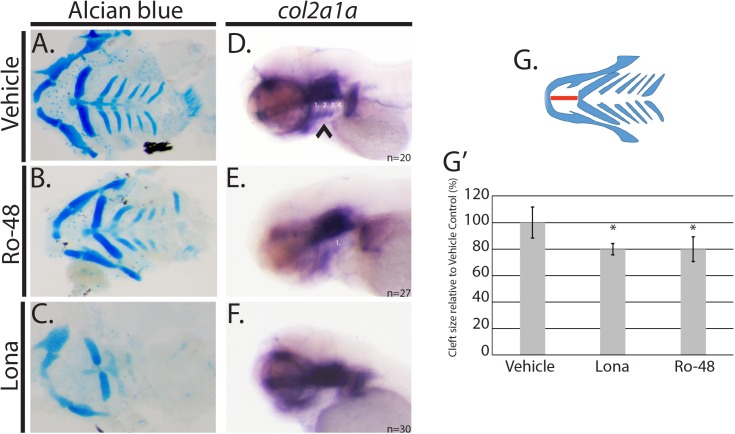
Fig 4. hmgcs1 regulates facial development by a cholesterol independent mechanism.
(A-C) Wildtype AB embryos were treated with 0.01% DMSO (vehicle), 8uM lonafarnib (Lona) or 3 uM Ro 48 8071 (Ro-48) beginning at 5 hours post fertilization and for a period of 4 days post fertilization (dpf). Alcian blue was used to analyze the cartilage structures of the developing zebrafish head and neck. (A-C) demonstrate the dissected viscerocranium of 4 day old larvae. (DMSO n = 30, Ro-48 n = 14, and Lona n = 8) (D-F) Whole mount in situ hybridization (ISH) was performed at 3 days post fertilization (protruding mouth stage) with embryos treated with 0.01% DMSO (vehicle), 8uM lonafarnib (Lona) or 2.5 uM Ro 48 8071 (Ro-48) (DMSO n = 20, Ro 48 n = 27, and Lona n = 30) (G) Schematic representation of viscerocranium. The extension of the Meckel’s cartilage (mandible) was measured as a detection for the presence of a facial cleft phenotype. Distance is indicated by the red line. (G’) Average was normalized and represented relative to the vehicle control group. Asterisk denotes statistical significance of p<0.001. Statistical analysis was performed using a T-test.

