Figure 5.
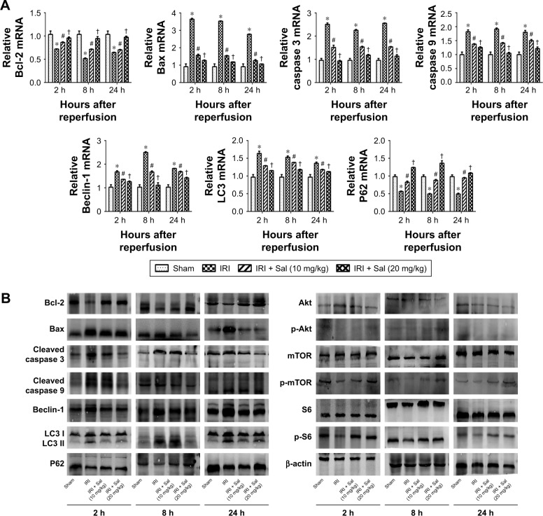
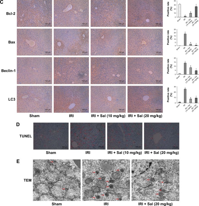
Sal pretreatment ameliorates apoptosis and autophagy in hepatic IRI.
Notes: (A) The relative mRNA levels of Bcl-2, Bax, caspase 3, caspase 9, Beclin-1, LC3, and P62. (B) Protein expression of apoptosis- and autophagy-related proteins. (C) Immunohistochemistry was used to detect Bcl-2, Bax, Beclin-1, and LC3 expression in liver tissues (original magnification, ×200). The ratio of brown area to total area was analyzed with Image-Pro Plus software 6.0. (D) TUNEL staining showed apoptotic cells (indicated by red arrows) in the four groups 8 hours after reperfusion (original magnification, ×200). (E) The autophagosomes were indicated by red arrows in TEM pictures. The results showed that there was more autophagosomes formation in the IRI group than in the Sal-treated group (original magnification, ×10,000). Data were given as mean ± SD (n=6, *P<0.05 for Sham versus IRI, #P<0.05 for IRI + Sal [10 mg/kg] versus IRI, and †P<0.05 for IRI + Sal [20 mg/kg] versus IRI + Sal [10 mg/kg]).
Abbreviations: IRI, ischemia–reperfusion injury; LC3, light chain 3; Sal, salidroside; TEM, transmission electron microscopy; TUNEL, terminal deoxynucleotidyl transferase dUTP nick end labeling.


