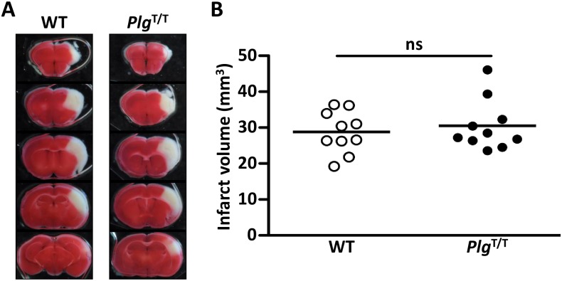
Fig 6. No exacerbation of PlgT/T mutation in the transient focal brain ischaemia model.
(A) Representative images of coronal sections of wild-type (WT) and PlgT/T mouse brains. Permanent occlusion of the distal M1 portion of the left middle cerebral artery and 15-min transient occlusion of the bilateral common carotid arteries were applied. After 24 hours, the brains were excised and stained with 2, 3, 5-triphenyl tetrazolium chloride. White areas represent brain infarction. (B) Infarct volumes. The infarct volume was adjusted for edema by dividing the volume by the edema index (left hemisphere volume / right hemisphere volume). No significant differences (p > 0.05) were observed between groups. Circles represent individual mouse data. Bars represent the mean values of groups.

